


The wide spread of osteoporosis in women in the postmenopausal period dictates the need to find new effective means of prevention and correction of pathological changes in bone tissue. The influence of peptide bio-regulators — cartilage preparation based on the extract from the cartilage tissue of calves and the medicine based on a short peptide — on the mineral density of bone tissue of rats in the experimental model of osteoporosis has been studied. As a result of the study, the osteoprotective effect of both studied substances has been established, while the effectiveness of the drug based on an extract from cartilage tissue was significantly higher. The substances had both a preventive effect on the state of bone tissue, preventing the reduction of bone mineral density after ovariectomy of rats, and corrective action, increasing the density of bone tissue reduced due to ovariectomy.
Postmenopausal osteoporosis is a key health problem in developed countries. Foreign experts consider that the disease has already taken the character of an epidemic. The medical and social significance of postmenopausal osteoporosis is due to its complications, first of all, fractures of the femoral neck and spine, which along with the deterioration of the quality of life significantly increase the mortality rate of women of older age groups. Despite a fairly wide Arsenal of pharmaceuticals that are used to treat systemic osteoporosis, the search for new tools that can effectively act the state of bone tissue, preventing its loss in the postmenopausal period continues. The results of the experimental study of peptide bio-regulators allow to predict the effectiveness of their use to improve metabolism in bone tissue .
Experimental models are an ideal tool for the study of pharmacological agents through their introduction into medical practice. It is caused by the fact that in the conditions of the experimental model the researcher can study molecular, cellular and systemic changes using all modern methods that cannot be studied in a clinical way. Certain requirements are imposed on experimental animals, among which the main ones are the ability of the model to accurately reproduce the studied pathological condition in humans; not to have undercurrent diseases; to be accessible, cheap and safe at work.
Rats fit better for the most of these rules in respect of postmenopausal bone loss. First, it is proved that the processes of bone loss in rats after ovariectomy reflect the main characteristics of postmenopausal osteoporosis in humans [5, 6]. Secondly, in rats, as in humans, after ovariectomy there is no tendency to complete restoration of bone tissue. Finally, rats are an informative model for the study of loss of both spongy and compact bone tissue which is especially important for the study of various types of osteoporosis, including postmenopausal one.
The purpose of the study: to examine the effect of peptide bio-regulators in different dosages and different application modes on the structural and functional state of bone tissue in experimental post ovariectomy models of osteoporosis in rats.
Material and methods of research
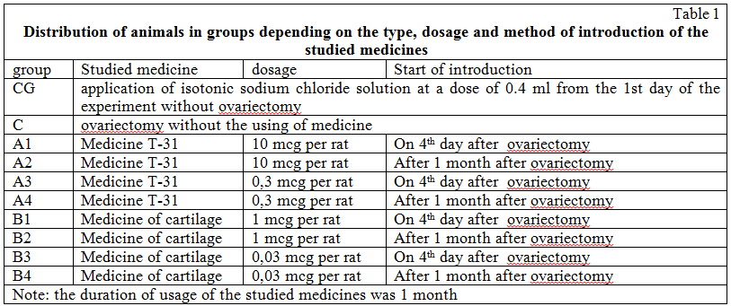
The effect of peptides on bone tissue in the experiment was studied in 100 Mature female rats type of Wistar with a body weight of 200-230 g at the age of 4-6 months, contained in the standard vivarium. The animals were divided into 10 groups per 10 individuals in each group, depending on the type, dosage and method of use of medicals. As the studied shot peptide, the cartilage preparation, which is an extract from the cartilage tissue of calves, and the medicine based on the short peptide were used. The studied preparations were administered to rats intramuscularly for 1 month: Cartilage preparation in doses of 1 mg and 0.03 mg per rat; medicine in doses of 10 µg and 0.3 µg per rat in 0.4 ml of sodium chloride physiological solution for modeling of postmenopausal osteoporosis rats were performed a bilateral ovariectomy. The median laparotomy was used as an operative access.
Rats were divided into groups per 10 animals in each of them: group A consisted of ovariectomized rats receiving injections of the medicine; group B — ovariectomized rats who were injected with cartilage; group C included ovariectomized rats who did not receive any medicine; control group consisted of non-operated animals receiving placebo in the form of 0.4 ml of physiological sodium chloride solution.
Depending on the scheme of medicine introduction, the following subgroups were established in these groups.
Rats in subgroups A1 received 10 mg per rat of the medicine per day during the first month after ovariectomy, animals of the subgroup A2 — 10 mg of the medicine per day during the second month after ovariectomy, animals of the subgroup A3 — 0.3 mg per day during the first month after ovariectomy, animals of the subgroup A4 — 0.3 mg per day during the second month after ovariectomy.
In group B subgroup B1 there were animals treated with 1 mg per rat for the medicine of cartilage per day during the first month after ovariectomy; subgroup B2 rats, which were injected 1 mg of cartilage per day during the second month after ovariectomy; subgroup B3 — 0,03 mg cartilage per day during the first month after ovariectomy; the subgroup B4 — 0,03 mg cartilage per day during the second month after ovariectomy.
The allocation of animals by groups is presented in Table 1.
Bone mineral density (BMD) was assessed using a two-photon x-ray densitometer «PRODIGY» (GE Medical systems, Lunar, model 8743, 2005), which has a software program «experimental animals», which allows to measure the mineral density and mineral saturation of bone tissue of the spine and the entire skeleton of small laboratory animals. Densitometry was performed three times: before the experiment, in 30 and 60 days after the experiment. The dynamics of indicators of BMD in absolute units and as a percentage have been studied.
The dynamics of mineral density in percent was calculated by the formula:
BMD(%)= [(BMD (after) -BMD (before) ) / BMD (before) ] x 100.
The obtained data were processed by the methods of variation statistics using the student’s aneurysm t-test related samples and monofactorial ANOVA analysis of variance.
The results of the study
Analysis of the results showed significant efficacy of cartilage medicine and peptide medicine in maximum dosages provided their use from the 30th day after ovariectomy (groups A2 and B2). In this method of application of peptides in 1 month after surgery, there was no significant increase in bone mineral density, which has been noted only in 2 months after ovariectomy. In the case of small doses of the studied medicines from the 30th day after the operation, there was a significant decrease in the mineral density of the skeleton after the end of taking the medicines compared with the initial indicators. Ovariectomy itself for 1 month on the dynamics of quantitative indicators of bone tissue is not affected, a decrease in BMD began to appear in 2 months after ovariectomy (group C).
Comparative dynamics of indicators of bone mineral density is presented in Table 2 and 3.
In the case of a monthly course of peptide bio-regulators taking from the 4th day after ovariectomy, the cartilage medicine in the maximum dosage (1 mg per rat) demonstrated the greatest efficiency. In its application, after 1 month, there was a significant increase of indicators; the achieved level of the indicators remained unchanged and after 2 months from the beginning of the experiment. The maximum dose of the medicine, introduced immediately after ovariectomy, a month later led to a significant increase in BMD. However, 1 month after discontinuation of the medicine (ie. after 2 months from the date of operation) there was a significant decrease in BMD compared to the baseline.
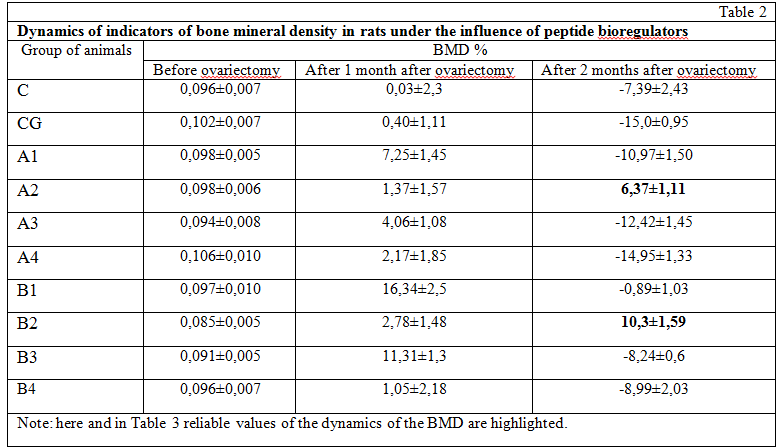
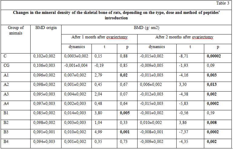
The monthly course of the medicine of cartilage and T-31 in small doses (0.03 mg and 0.3 mg per rat, respectively), started from the 3rd day after ovariectomy, did not lead to an improvement in bone tissue parameters. A month after its completion, there was a significant decrease in BMD compared to the values at the time of the experiment.
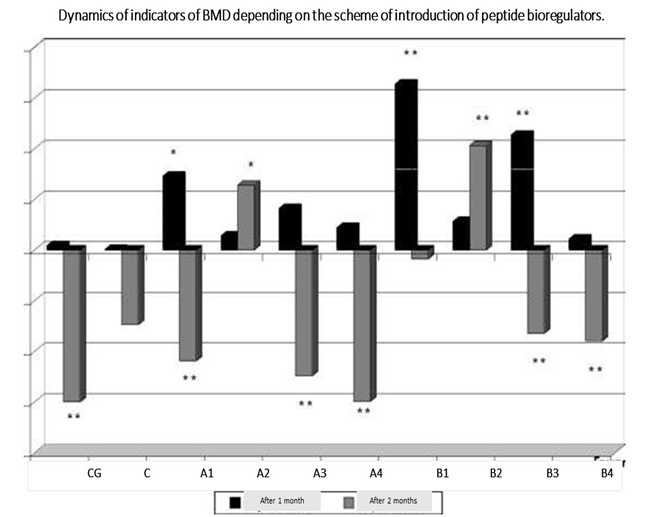
The proved dependence of the dynamics of BMD on the treatment method was demonstrated by the ANOVA dispersion method (figure).
Summary
- To prevent the loss of bone mass in ovariectomized rats, the most effective is the use of the medicine based on the extract of cartilage tissue in animals in large doses (1 mg per rat) 1 month after ovariectomy, reliably increase in a month BMD compared to baseline. After discontinuation of the medicine, the BMD values decrease to the initial level within a month. For a more accurate determination of the duration of the osteo protective effect of the cartilage preparation in this dosage, further studies are required.
- The use of medicine T-31 in the maximum dose (10 mg per rat) also significantly improves skeletal BMD, but after discontinuation of the medicine, its reduction is observed, which suggests the need for continuous use of this medicine.
- Small doses of peptides were effective only in the case of cartilage immediately after ovariectomy, which allowed to significantly improve the structural and functional state of the bone tissue of experimental animals, but only at the time of introduction of the medicine, which also suggests the need for continuous introduction of them latter.