


At the present time the study of the peptides’ properties [Dudgeon W. D. et others, 2016] has a high interest. Peptides have the same structure as proteins (proteins), but the size of these molecules is smaller. It is also important to note that short peptides, being a natural metabolic product present in the body, cannot be identified in blood or urine. In this case the study of the properties of individual structures on cell cultures can only be provided.
Peptide IPH-AEN contains a low molecular weight peptide, has chondroprotective and osteoprotective properties and a normalizing effect on cartilage and bone tissue.
Experimental studies have shown that the peptide IPH-AEN regulates metabolic processes in chondrocytes and osteocytes, increases the reserve capacity of the body, which suggests the effectiveness of the peptide IPH-AEN to normalize the functions and restore the cartilaginous and skeletal systems in human disorders of various origins.
Thus, the aim of the present research was to study chondroprotective, osteoprotective and other properties of the peptide.
Study design
1 Phase.
To examine the cytostatic and onco-protective properties of the peptide IPH-AEN in relation to the bone and cartilaginous system, have been chosen embryonic stem cells (ESCs), that refer to pluripotent type, which means that they can be differentiated into all three primary germ layers: ectoderm, endoderm and mesoderm, from which the cartilage system tissues are formed in the future. The human embryo reaches the blastocyst stage, from which stem cells are obtained on days 5-6 after fertilization.
The genes responsible for the formation of bone and cartilage are the COMP gene (Transcript: NM_000095) and the COL2A1 gene.
Mutations in the cartilage of thrombospondin (COMP) cause achondroplasia and pseudo epiphyseal dysplasia, accompanied by the defeat of the large joints. In connection with these data, it has been decided to study the activity of this gene in the application of the IPH-AEN peptide.
Gene mutations and changes in COL2A1 gene expression are associated with the severity of degenerative lesions of bone and cartilage. The COL1А1 gene encodes the alpha-1 protein of the type I collagen chain and is potentially significant in the formation of a genetic predisposition to osteoporosis and bone fragility at risk of fractures. Thus, it also has been decided to study the activity of this gene in the application of the IPH-AEN peptide.
We evaluated the expression of genes responsible for the ontogenesis of bone and cartilage tissue.
2 Phase.
The second stage of the present experimental study was the evaluation of marker biological active molecules by immune fluorescent method using primary antibodies to MMP13 (1:120, Abcam) and p53 protein (1:50, Abcam).
We have selected the following marker biologically active molecules:
1) Protein MMR13 is a marker of remodeling of intercellular matrix and functional activity of chondrocytes. Matrix metalloproteinases (MMP), belonging to the family of zinc metalloproteinases, participate in the exchange of intercellular matrix proteins. The leading role in the process of physiological remodeling of hyaline cartilage of the joint is assigned to metalloproteinases belonging to the class of collagenases (MMP-1, MMP-13, etc.). Normally, metalloproteinases are secreted by fibroblasts, chondrocytes, epithelial cells, macrophages in very small amounts in the inactive state. An important feature of metalloproteinases is their ability to induction under the influence of various factors. Regulation of collagenase activity is carried out by cytokines, growth factors, various chemical compounds (bacterial lipopolysaccharides etc.), factors affecting the cell membrane. MMP activity is suppressed by tissue inhibitors of metalloproteinases (TIMP), as well as hormones (glucocorticosteroids, etc.). Earlier it was considered that the main role in the destruction of the matrix belongs to MMP-1, then it was established and the important role of MMP-13, which destroys collagen type II, stromelyzin, etc. It is known that in the activation mechanisms of metalloproteinases the significant role is given to signal proteins S100A8 and A9. As a result, NF-kB receptors of synovial fibroblasts are activated, which determines the release of many cytokines, such as IL-6, IL-10, GM-CSF, IL-8 and chemoattractant in monocytes, which, in turn, plays a significant role in the activation of metalloproteinases and causes their high activity in osteoarthrosis [Demkin S. A. et al., 2017].
2) The aging of polypotent cells in culture is associated with increased activity of the p53 gene. In mice with mutation in the p53 gene, there was increased resistance to the development of tumors, combined with a reduction in life expectancy. P53 protein plays a key role in endogenous antitumor mechanisms [Donekover et al., 2002]. P53 protein controls the course of cell cycle processes, as well as the absence of damage in the genome that could lead to further development of pathology. Protein p53 is a transcriptional factor that serves as a suppressor of malignant tumors by activating apoptosis in the tissues of the body. The p53 protein is activated when DNA is damaged, as well as when stimulation can lead to such damage or are a signal of cell aging and a disorder of its functional activity [Arshad H. et al., 2010]. P53-dependent apoptosis avoids the accumulation of mutations, and, in case when they have already occurred, p53-dependent apoptosis allows to eliminate such potentially dangerous cells [Burtis C., Ashwood E., Bruns D., 2006].
Research
To study of the peptide IPH-AEN properties, we used the following cell cultures of the Russian collection of vertebrate cell cultures (RCVCC):
SC5
Origin: human, embryonic stem cells (ESCs), blastocyst (5-6 days of growing), obtained as a result of IVF.
Science. 1998. 282: 1145 – 1147; Ontogenesis. 2011. 42 (4): 249 – 263;
Cytology. 2012. 54 (1): 5 – 16.
Morphology: colonies of rounded cells with high nuclear/cytoplasmic ratio.
Method of cultivation: monolayer; colonies attached to mitotic inactivated (mitomycin-C) feeder layer of mesenchymal cells of the bone marrow of the human embryo.
Cultivation conditions: medium — Knockout Dulbecco’s modified Eagles medium serum — Knockout Serum Replacement 20% other ingredients – NEAA 1%, L-glutamine 2mM, 2 — mercaptoethanol 0.1 mM, bFGF — 8 ng/ml
the procedure of replanting – mechanical replanting of ESC culture was carried out under the control of a microscope by dividing the colony into fragments using a disposable scalpel and transferring them to a new layer of the feeder; daily change of environment, replanting every 5-6 days.
cryopreservation — growth medium, 10% DMSO, 5×10⁵ cells/ml in ampoule.
Viability after cryopreservation: 60 % (painted trypan blue on the zero passage)
Control of contamination: bacteria, fungus and Mycoplasma were not detected.
Control of the identity of the species: karyological analysis.
Karyology: 2n= 46, modal chromosome number 46 (98.0+0.9 %), normal human karyotype (46, XX), number of polyploids (0.2 + 0.2%).
DNA profile (STR): Amelogenin: X, X
CSF1PO: 12, 13
D13S317: 8, 11
D16S539: 9, 12
D5S818: 9, 11
D7S820: 8,10,12
THO1: 6, 9.3
TPOX: 10, 11
vWA: 17,17
Other characteristic:
The average time of one cell population doubling is 28.2 hours.
Immortalized line made more than 120 doubling the cell population.
Expression of surface antigens typical for human ESCs: SSEA-4, TRA-1-60 and transcription factors Oct-4, Nanog.
The ability to spontaneously differentiate into derivatives of 3 germ leaves.
Ability to form in vivo teratos containing derivatives of 3 germinal layer.
Field of application: cell biology, embryology.
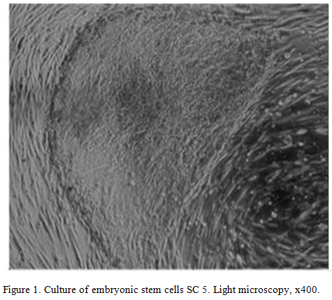
Collections: Institute of Cytology of the Russian Academy of Sciences (figure 1) (Federal state budgetary institution of science «Institute of Cytology of the Russian Academy of Sciences, M.S. Bogdanova, G.G. Polyanskaya, A.M. Koltsova, «Cell cultures» Information Bulletin. Issue.34 (2018))
Method of research
Group for the study:
Group 1 – the measurement of the expression of molecules before the study,
Group 2 — control (addition of nutrient medium, serum albumin incubation),
Group 3 – addition of a control peptide of Glu-Trp dipeptide at the concentration of 100 micrograms (µg);
Group 4 – addition of IPH-AEN peptide at the concentration of 100 micrograms (µg).
The study used peptides IPH-AEN and Glu-Trp in the form of a lyophilized powder, which were dissolved with sterile water for injection in a volume of 10 ml to a final peptide concentration of 100 µg.
As previously shown for most dissociated cell cultures the most effective concentration is the one of peptides 100 µg/ml according to long-term experience of peptides [Linkova N.S. et al., 2016; Khavinson V. et al., 2017].
As a control, selected immune-protective peptide Glu-Trp with the known and well described in the literature properties [Morozov V.G., Khavinson V.Kh., Malinin V.V., 2000, Khavinson V.Kh., Morozov V.G., 2001, Khavinson V.Kh., Kuznik B.I., Linkova N.S., Pronyaeva V.E., 2013].
PCR-method with the use of own primers and Novocasta reagents and sets of monoclonal antibodies of Biosource company (Belgium). were used to measure the level of gene expression.
Cell smears were treated with appropriate primary antibodies according to the standard Protocol:
- Triple rinsing with phosphate buffer saline- PBS(рН=7,2) for 5 minutes;
- Permeabilization of cells 0,1% Triton X-100, dissolved in PBS during for 15 minutes;
- Rinsing 3 times in PBS (per 5 minutes);
- Incubation in 1% bovine serum albumin (dissolved by PBS, pH 7.5) for 30 minutes to block nonspecific binding;
- Incubation with primary antibodies, 60 minutes;
- Rinsing 3 times in PBS (per 5 minutes);
- Incubation with secondary antibodies conjugated with fluorochrome Alexa Fluor 488 (1:1000, Abcam) for 30 minutes at indoor temperature in the dark;
- Rinsing 3 times in PBS (per 5 minutes);
- Drawing cell cores with pigment Hoechst 33258 (Sigma, USA) (1:100 from stock solution in dH2O) for 1 minute (the pigment is used as a fluorescent marker DNA, when binding to which its fluorescence is enhanced).
- Rinsing in PBS (per 5 minutes);
- Custody of prepared specimens under cover glasses in the installer environment Dako Fluorescent Mounting Medium (Dako).
The study of specimens was carried out in a confocal microscope Olympus FluoView FV1000 at zoom of 200, 400, 600. Blue color fluoresces the expression of the studied marker. Measurement of the relative areas of expression has been conducted in %. The relative area of expression was calculated as the ratio of the area occupied by immune- positive cells to the total area of cells in the field of view and expressed as a percentage. The dynamics of gene expression is measured in conventional units, the base level is taken as 10.
Statistical processing of results
Statistical processing of experimental data included calculation of arithmetic mean, standard deviation and confidence interval for each sample and was carried out in Statistica 11.0. If the data were normally distributed, the differences in means were determined by the Student test (t).
Research results and discussion
Thus, in figure 2 has shown that the peptide IPH-AEN significantly increases the expression of the gene COMP, responsible for the normal formation of bone and cartilage tissue, in particular, regulating metabolism in bone and cartilage tissue and provide protective actions against joint damage in humans, so, the peptide IPH-AEN counteracts degenerative processes in the cartilage tissue of the joints thereby providing prevention of joint diseases.
Figure 3 it has been shown that the peptide IPH-AEN significantly increases the expression of the gene COL2A1, controlling degenerative lesions of cartilage, in particular, significant in the formation of genetic predisposition to osteoporosis and bone fragility with the risk of fractures.
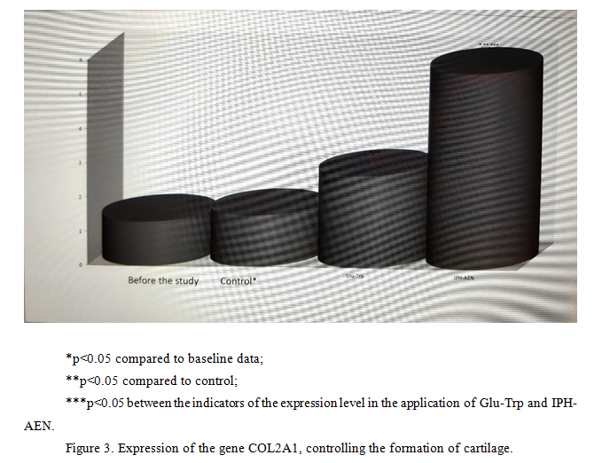
Thus, from the above data it can be seen that under the influence of the peptide IPH-AEN in human cell culture there is a statistical
ly significant increase in the expression of genes responsible for the ontogenesis of bone and cartilage tissue, namely the synthesis of cartilage cells, which provides rapid regeneration of bone tissue, improves the production of collagen and hyaluronic acid. These data show that the peptide IPH-AEN significantly increases in human cell culture «cascade» of signal molecules, which is necessary to activate the processes of proliferation and differentiation of stem cells in the cells of cartilage, the formation of cartilage systems, regulation of metabolism in cartilage tissue and providing protective properties against joint damage, control of degenerative lesions of cartilage tissue, osteoporosis and bone fragility with the risk of fractures in humans.
The effect of the peptide IPH-AEN on ММР13 protein expression in cultures of human cells
Figure 4 shows that the use of the peptide IPH-AEN increases the expression of MMP13 in 9.6 times from the initial level, which is a key factor in the remodeling of the intercellular matrix and functional activity of chondrocytes, thereby regulating the synthesis of cartilage cells and providing rapid regeneration of bone tissue.
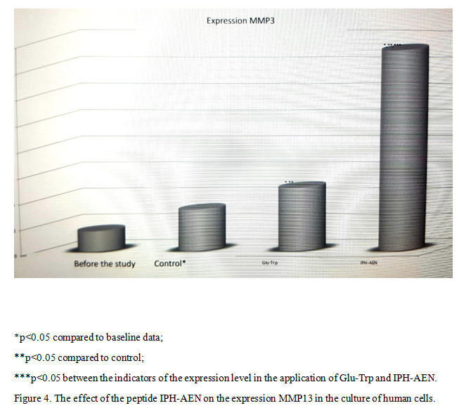
Thus, the use of the peptide IPH-AEN has chondroprotective and osteoprotective character, in particular, induces differentiation of polypotent chondrogenic and osteogenic cells in the direction of normal cartilage tissue formation and its regeneration. In particular, these data indicate that the use of the peptide IPH-AEN provides synthesis of cartilage cells and rapid bone regeneration, which leads to the maintenance of full functionality of the joints.
The effect of the peptide IPH-AEN on the expression of p53 protein in cultures of human cells
Figure 5 shows that the use of the peptide IPH-AEN increases the protein p53 production, which is a transcriptional factor that serves as a suppressor of malignant tumors by activating apoptosis in the tissues of the body, which leads to make a conclusion about the antitumor properties of the studied peptide.
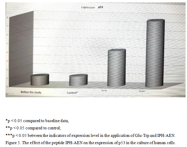
P53-dependent apoptosis also avoids the accumulation of mutations, and, in the case where they have already arisen, p53-dependent apoptosis allows to eliminate such potentially dangerous cells for the body, which leads to make a conclusion about the cytoprotective effect of the studied peptide.
The data indicate a high Onco-protective activity of the peptide IPH-AEN in respect of cells of a bone-cartilage system according to the expression of biological molecules in cell culture.
Conclusion
The performed studies confirm the high biological activity of the peptide IPH-AEN in relation to the normal formation of bone and cartilage tissue, regulation of metabolism in cartilage tissue and the provision of a protective action against joint damage in humans, as well as control of the development of degenerative lesions of cartilage tissue, in particular, with osteoporosis and injuries. Peptide IPH-AEN counteracts degenerative processes in the cartilage of the joints, thereby providing prevention of joint diseases.
The data obtained show that under the influence of the peptide IPH-AEN in the culture of human cells there is a statistically significant increase in the expression of genes responsible for the ontogenesis of bone and cartilage tissue, namely the synthesis of cartilage cells, which provides rapid regeneration of bone tissue, improves the production of collagen and hyaluronic acid. These data show that the peptide IPH-AEN significantly increases in human cell culture «cascade» of signal molecules, which is necessary to activate the processes of proliferation and differentiation of stem cells in the cells of cartilage, the formation of cartilage systems, regulation of metabolism in cartilage tissue and providing protective properties against joint damage, control of degenerative lesions of cartilage tissue, osteoporosis and bone fragility with the risk of fractures in humans.
The use of the peptide IPH-AEN has chondro-protective and osteoprotective character, in particular, induces differentiation of polypotent chondrogenic and osteogenic cells in the direction of normal formation of cartilage tissue and its regeneration. In particular, these data indicate that the use of the peptide IPH-AEN provides synthesis of cartilage cells and rapid bone regeneration, which leads to the maintenance of full functionality of the joints.
The data indicate a high onco-protective activity of the peptide IPH-AEN in relation to the cells of the cartilage according to the expression of biological molecules in cell culture.
Literature
- Zabello T.V., Miromanov A.M., Mirimanova A.N. Genetic aspects of osteoarthritis // Fundamental research – 2015. – № 1-9. – p. 1970-1976
- Linkova N.S., Drobantseva A.O., Orlova O.A., Kuznetsova E.P., Polyakova O.V., Kvetnoy I. M., Vinson V.Kh. Peptide regulation of functions of skin fibroblasts upon aging in vitro // Cell technologies in biology and medicine. – 2016. — №1. – p. 40-44.
- Mutovin G.R. Fundamentals of clinical genetics (genomics and proteomics of hereditary pathology). Textbook for universities in 2 volumes. Issue. 3. M.: GEOTAR-media 2008.
- Khavinson V. H. Peptide regulation of aging. SPb.: Science,2009. — 50 p.
- Arshad H., Ahmad Z., Hasan S.H. Gliomas: correlation of histologic grade, Ki67 and p53 expression with patient survival // Asian Pac J Cancer Prev. – 2010. – Vol. 11. – N 6. – P. 1637-1640;
- Meienberg J, Rohrbach M, Neuenschwander S, Spanaus K, Giunta C, Alonso S, Arnold E, Henggeler C, Regenass S, Patrignani A, Azzarello-Burri S, Steiner B, Nygren AO, Carrel T, Steinmann B, Mátyás G. Hemizygous deletion of COL3A1, COL5A2, and MSTN causes a complex phenotype with aortic dissection: a lesson for and from true haploinsufficiency. Eur J Hum Genet. 2010;18:1315–21.
- Trzeciak T. MicroRNAs: Important Epigenetic Regulators in Osteoarthritis / T. Trzeciak, M. Czarny-Ratajczak // Curr. Genomics. – 2014. – № 6. – P. 481–484.
- Tsezou A. Osteoarthritis year in review 2014: genetics and genomics // Osteoarthritis Cartilage. – 2014. – № 12. – P. 2017–2024.