Introduction
At the present time the study of the peptides’ properties [Dudgeon W. D. et others, 2016] has a high interest. Peptides have the same structure as proteins, but the size of these molecules is smaller. It is also important to note that short peptides, being a natural metabolic product present in the body, cannot be identified in blood or urine. In this case the study of the properties of individual structures on cell cultures can only be provided.
Peptide IPH-AEN contains a low molecular weight peptide, has chondroprotective and osteoprotective properties and a normalizing effect on cartilage and bone tissue.
Experimental studies have shown that the peptide IPH-AEN regulates metabolic processes in chondrocytes and osteocytes, increases the reserve capacity of the body, which suggests the effectiveness of the peptide IPH-AEN to normalize the functions and restore the cartilaginous and skeletal systems in human disorders of various origins.
Thus, the aim of the present research was to study chondroprotective, osteoprotective and other properties of the peptide.
Characteristics of the experiment
The most frequently used type of laboratory animals for peptide properties studies, recommended by the Ministry of health of the Russian Federation in the Manual for preclinical studies of drugs [Mironov A. N., Bunatyan N. D. et al., 2012], are rats, as pharmacokinetic processes are similar to those in humans.
To study the properties of the peptide IPH-AEN, has been created a model of acetabulum damage. Obtaining a model of linear through bone and cartilage damage of the acetabulum (RF patent No. 2470378) was performed under sterile conditions under General combined anesthesia: zoletil 100 («Virbac Sante Animale», France) 8 mg/kg, xylazine 2 % («Alfasan International B.V.», Holland) 4.0 mg/kg, intramuscularly. In the area of the femoral neck, the joint capsule was opened and the head dislocated from the acetabulum. Further, by surgical saw 0.2 mm thick, a transverse cut of the acetabulum was made from the side of its dorsal part to the level of the middle of the articular fossa, after which the head and neck of the femur were resected.
We studied 40 rats aged 13.8±1.2 months and weighing 423.1±7.5 g, which created the conditions of muscle injury.
Daylight was 12 hours. For food animals used complete feed for rodents with additional feeding in the form of fruits and vegetables. Water was used by animals independently from drinkers. Food and liquid were taken by animals ad libitum. Current cleaning of cells was carried out daily. General cleaning with disinfection of cells was performed weekly. All procedures of keeping animals, manipulations and testing of the data were carried out in accordance with ISO 10993-1-2003 and state standard RISO 10993.2-2006.
Rats were divided into 2 groups – control (n=20) and main (n=20). Rats of the main group orally using the pipette, allowing to control the amount and consumption of the liquid, was introduced a solution consisting of water for injection at a dosage of 1 ml in which dissolved lyophilized powder peptides IPH-FH at a concentration of 0.60 micrograms (mcg) per body weight of rat per day (minimum dosage at which there were signs of improvement after the application of peptides) for 14 days. The drug was administered per os directly into the oral cavity under supervision and tracking its ingestion.
After 14 days, the rats were killed, then the joint capsule was removed, fixed by immersion in a solution of 4% paraformaldehyde in a phosphate buffer (PBS pH = 7.3) for 24 hours at a temperature of 4 ° C. Produced slices with a thickness of 20 µm using cryotome of Leica CM 1510S model (Germany). The sections were then mounted on a slide and stained with hematoxilin and eosin.
For the study we used the microscope Olympus IX81. The microscope was equipped with a digital camera Olympus DP72 (Japan), connected to a personal computer. Photo and video recording of technological processes (experimental processes) with animals was not carried out in accordance with the principles of biomedical ethics and due to the lack of permission of the ethical Committee.
Statistical processing
To assess the reliability of the difference in the results obtained in the groups before the use of drugs, compared with the groups after the use of drugs, the Dunnet criterion was used. If the data were normally distributed, the differences in means were determined by the Student test (t).
Result of the study
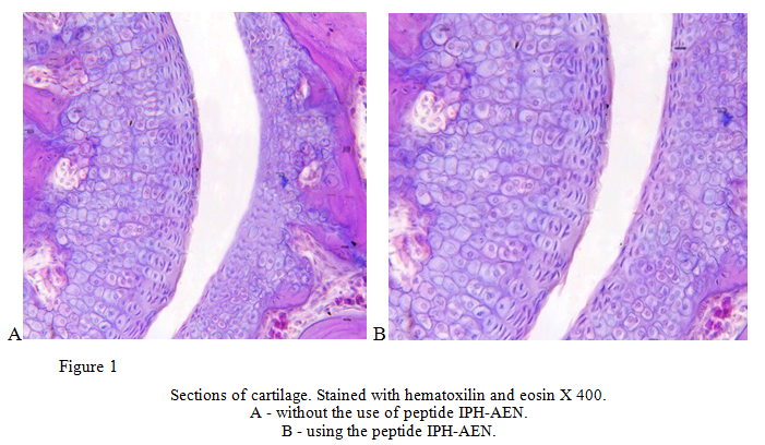
During the experiment it was shown that the use of the peptide IPH-AEN significantly increases the reparative properties of cartilage. When using this peptide, it was noted that the zone of damage was filled with granulation tissue with a large number of thin-walled capillaries lined from the inside with flattened nucleated endothelial cells. On the surface of the injured bone trabeculae there were active osteoblasts, few osteoclasts were found. In the bone tissue of the fragments near the line of damage, nucleated osteocytes were determined, on the periosteal and endosteal surfaces – active osteoblasts. While in control group it was found that the diastasis between atomtime was filled with fibrin and tissue detritus, including fields changed necrotic cells with a pronounced criminosa, karyorhexis or karyolysis. The cell composition was dominated by cells characteristic of the inflammatory stage of the reparative process — segmental leukocytes and mononuclear phagocytes (figure 1).

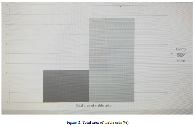
During the experiment we revealed the total area of viable cells (figure 2).

The use of the peptide IPH-AEN increases the total area of viable cells by 2.7 times. These data confirm the fact that the use of the peptide IPH-AEN helps to improve the regeneration and restoration of bone and cartilage tissue, the synthesis of cartilage cells, which provides rapid regeneration of bone tissue, improves the production of collagen and hyaluronic acid after damage on the experimental model.
Conclusion
Thus, the use of the peptide IPH-AEN improves repair and regeneration of bone and cartilage tissue after damage, reduces the degree of inflammatory reaction, in other words, the peptide IPH-AEN has a high reparative and regenerative activity against bone and cartilage tissue and has anti-inflammatory properties, and also counteracts degenerative processes in the cartilaginous tissue of the joints thereby providing prevention of joint diseases.
Literature
- Zabello T. V., Miromanov A. M., Mirimanova N. A. Genetic aspects of osteoarthritis // Fundamental research. – 2015. – № 1-9. – p. 1970-1976
- Linkova N. S., Drobantseva A. O., Orlova O. A., Kuznetsova E. P., Polyakova O. V., Kvetnoy I. M., Vinson V. Kh. Peptide regulation of functions of skin fibroblasts upon aging in vitro // Cell technologies in biology and medicine. – 2016. — №1. – P. 40-44.
- Khavinson V. Kh. Peptide regulation of aging. SPb.: Science, 2009. — 50 p.
4.Arshad H., Ahmad Z., Hasan S.H. Gliomas: correlation of histologic grade, Ki67 and p53 expression with patient survival // Asian Pac J Cancer Prev. – 2010. – Vol. 11. – N 6. – P. 1637-1640;
5.Trzeciak T. MicroRNAs: Important Epigenetic Regulators in Osteoarthritis / T. Trzeciak, M. Czarny-Ratajczak // Curr. Genomics. – 2014. – № 6. – P. 481–484.
6.Tsezou A. Osteoarthritis year in review 2014: genetics and genomics // Osteoarthritis Cartilage. – 2014. – № 12. – P. 2017–2024.


