


For the last year the number of cases of osteoporosis has recently been increased in postmenopausal women. Such process makes it
necessary to obtain new effective tools that could correct and prevent pathological changes in the bone tissue. An experimental model of the
development of osteoporosis was carried out, taking into account the use of peptide bioregulators based on short peptides that affected the
bone mineral density in rats. During the study, these peptides had a significant osteoprotective effect with high efficiency. Due to the corrective
action, the bone density increased. Due to the preventive action, a decrease in bone mineral density was prevented after ovariectomy in rats.
In recent years, osteoporosis in postmenopausal women has been one of the key health problems in developed countries. Many experts believe that such a trend today has a huge scale. The medical problem is mainly related to the complications that the disease causes. So significant cases occur in women and are associated with fractures of the spine and femoral neck. We can say that such consequences lead to an increase in mortality, and the quality of life in healthy people is significantly reduced. [1, 8]. There are a significant number of medicals for the treatment of systemic osteoporosis, however, the search for new and more effective means to prevent bone loss in the postmenopausal period is urgent. An experimental study using peptide bioregulators showed efficiency in the metabolism of bone tissue. [3].
This experimental model was carried out taking into account the study of characteristics at the molecular, cellular level, and systemic changes were also investigated. This model will allow us to further implement the obtained data in clinical studies and introduce them into clinical studies. [2]. For the experimental model, rats were taken like the most accurately reproduce the pathological state of a person. The animals had no hidden diseases, and are the most accessible for research and safe to work with. [7, 10].
It is proved that the processes of bone loss in rats after ovariectomy are similar to the main characteristics of the process of postmenopausal osteoporosis in humans [5, 6]. Also, in rats, as in humans, there is no process for the complete restoration of bone tissue after a behavioral ovariectomy. [4]. Rats are also able to show how spongy and compact bone tissue is lost. [9, 11] The last point is an important indicator in the study of types of osteoporosis, including in postmenopausal women.
The main object of the current study was to provide experimental model to examine the effect of peptide bio-regulators, including short peptides, in different dose and usage modes on the structural and functional state of bone tissue of osteoporosis in rats after ovariectomy.
Material and methods of research
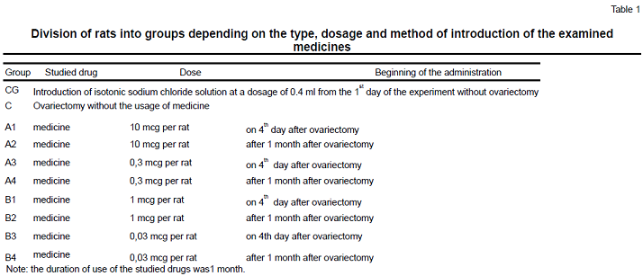
To study the effect of peptides on bone tissue, 100 mature female Wistar rats with a body weight of 200-230 g at the age of 4-6 months were taken. The rats were kept in a standard vivarium. The animals were divided into 10 groups of 10 individuals in each group, which were divided according to the type, doses and methods of medicines administration. The studied peptide was an injection from a cartilage preparation, which is an extract from the cartilage tissue of calves, and drugs based on a short peptide. The studied drug was administered intramuscularly to rats for 1 month: a cartilage preparation in doses of 1 mg and 0.03 mg per rat; a drug in doses of 10 mcg and 0.3 mcg per rat in 0.4 ml of sodium chloride saline solution. To create a process close to modeling postmenopausal osteoporosis, rats underwent a bilateral ovariectomy. An operative access was made by the median laparotomy.
All rats for research have been divided into some groups per 10 animals in each of them: group A consisted of ovariectomized rats receiving injections of the medicine in doses of 1 mg and 0.03 mg per rat; group B — ovariectomized rats who were injected with cartilage in doses of 10 μg and 0.3 μg per rat; group C included ovariectomized rats who did not receive any medicine; control group consisted of non-operated animals receiving placebo in the form of 0.4 ml of physiological sodium chloride solution
Inside the abovementioned groups have been selected subgroups which depending on the way of introduction the medicine. There have been established 4 subgroups.
Rats in subgroups A1 have got 10 mg per rat of the medicine per day during the first month after ovariectomy, rats of the subgroup A2 have received 10 mg of the medicine per day during the second month after ovariectomy, rats of the subgroup A3 have received 0.03 mg per day during the first month after ovariectomy, rats of the subgroup A4 — 0.03 mg per day during the second month after ovariectomy.
In group B subgroup B1 there were animals treated with 1 mg per rat for the medicine of cartilage per day during the first month after ovariectomy; subgroup B2 rats, which were injected 1 mg of cartilage per day during the second month after ovariectomy; subgroup B3 — 0,03 mg cartilage per day during the first month after ovariectomy; the subgroup B4 have received 0,03 mg cartilage per day during the second month after ovariectomy. The division of rats by groups is illustrated in Table 1.
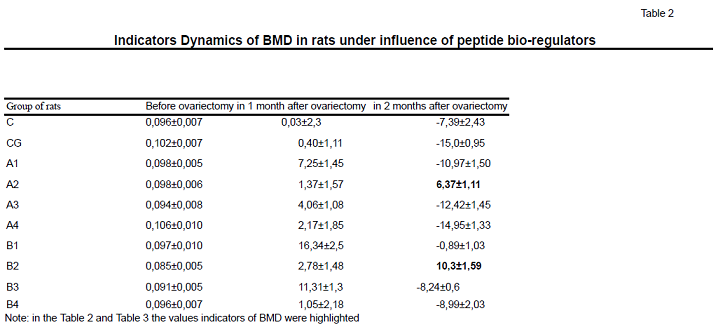
Bone mineral density (BMD) was examined by a two-photon x-ray densitometer «PRODIGY» (GE Medical systems, Lunar, model 8743, 2005), which has have a software program «experimental animals», which allows to measure the mineral density and mineral saturation of bone tissue of the spine and the entire skeleton of small laboratory animals. Densitometry has been used three times during the study: first time it was used before the experiment, the second time it was performed in 30 days after the experiment, and the last time it was used in 60 days after the experiment. The dynamics of indicators of BMD in absolute units and as a percentage have been studied.
The dynamics of mineral density in percent was calculated by the formula:
BMD(%)= [(BMD (after) -BMD (before) ) / BMD (before) ] x 100.
The obtained data were processed using the methods of variation statistics, so called, the Student’s t-test related samples and monofactorial ANOVA analysis of variance.
Results of the study
After receiving of the results of the study, it was shown significant efficacy of cartilage drug and peptide medicine in maximum dosages used after the 30th day after ovariectomy (groups A2 and B2). Such way of peptides introduction during just 1 month after surgery has had no significant increase in bone mineral density, instead of indicators after 2 2 months after ovariectomy. After 2 months the indicators has shown high effective. The method of small doses indicatoins of the examined medicines after 30 days after the operation, has not shown a significant decrease in the mineral density of the skeleton after the end of taking the medicines compared with the initial indicators. Ovariectomy itself has not affected in case of the dynamics of quantitative indicators of bone tissue for the first month, a decrease in BMD started to appear only in 2 months after ovariectomy (group C).
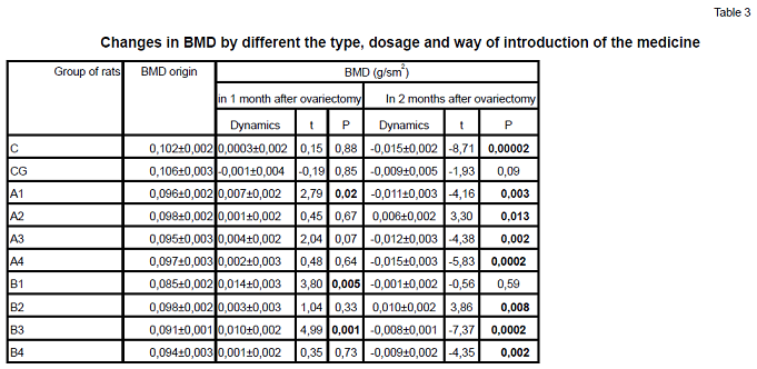
Comparative dynamics of indicators of bone mineral density is shown in Table 2 and 3. During a monthly course of peptide bioregulators injection from the 4th day after ovariectomy, the cartilage medicine in the maximum dosage (1 mg per rat) has shown the greatest efficiency. After 1 month of the injections, a significant increase of examined indicators has been noted; after that this level remained unchanged even after 2 months from the start of the experiment. The maximum dose of the medicine, which was injected immediately after ovariectomy, has shown a significant increase in BMD in month after start of introduction. However, 1 month after stop of introduction of the medicine (ie. after 2 months from the date of operation) there was noted a significant decrease in BMD compared to the baseline.
The monthly course of the medicine in small doses (0.03 mg and 0.3 mg per rat, respectively), which started from the 3rd day after ovariectomy, has not lead to an significent in bone tissue indicators. In a month after its finish, there was noted a significant decrease in BMD compared to the values at the time during the experiment. The proved dependence of the indicators of BMD on the treatment way has been shown by the ANOVA dispersion method (figure).
Conclusion
1. To decrease the loss of bone mass in ovariectomized rats, the most effective way was used medicine on the extract of cartilage issue in rats in large doses (1 mg per rat) during 1st month after ovariectomy.Such method certainly increase in a month BMD compared to baseline. After discontinuation of the medicine, the BMD has decreased to the original level within a month. For a more certain time of application peptide medicine is needed the further studies to receive the osteo protective effect in this dosage.
2. The application of the medicine in the maximum dose (10 mg per rat) also provide an improve in skeletal BMD, however, after discontinuation of the medicine, its reduction has noted, which suggests the need for further medicine introduction.
3. Introduction of peptides in small doses has had more effective results only in the case of cartilage immediately after ovariectomy, which allowed to significantly improve the functional and structural state of the bone tissue in experimental rats, but only during the time of introduction of the medicine, which also suggests the need for further application of the medicine.
Литература
1. Povoznyuk V., Grigoryeva N. Menopausa and musculoskeletal system.— 2004.—512 p.
2. Frolkis V., Povoznyuk V., Yevtushenko O. Experimental osteoporosis (models, mechanism of development of age-related osteoporosis. 2 vol. Т.1. — 2004.—p. 356-388.
3. Khavinson V., Anisimov V. Peptide bioregulators and aging. Saint-Petersbourg: 2003.—223 p.
4. Abe T, Chow JWM, Lean JM, Chambers TJ. Estrogen does not restore bone lost after ovariectomy in the rat // J Bone Miner Res. — 1993.—Vol. 8.—P 831-838.
5. Aerssenss J, Boonen S, Lowet G, Dequeker J. Interspecies difference in bone composition, density and quality: Potential implications for in vivo bone research // Endocrinology.—1998.— 139.—P 663-670.
6. Barlet JP, Coxam V, Davicco MJ, Gaumet N. Modeles ani- maux d’osteoporose post-menopausique // Reprod Nutr Rev.—1994. —34.—P. 221-236.
7. Davidson MK, Lindsey JR, Davis JK. Requirements and selection of an animal model // Isr J Med Sci.—1987.—23.—P. 551555.
8. Marcus R, Wong M, Heath H, Stock J. Antiresorptive Treatment of Postmenopausal Osteoporosis : Comparison of Study Designs and Outcomes in Large Clinical Trials With Fracture as an Endpoint // Endocrine Reviews.—2002.—Vol. 23 (1).— P. 16-37.
9. Sietsma WK. Animal models of cortical porosity // Bone.-1995. —Vol. 17.—P 297-305.
10. Simon AT. Animal models of osteoporosis — necessity and limitations // Europian Cells and Materials.—2001.— Vol. 1.—P 66-81.
11. Wronski TJ, Yen CF. The ovariectomised rat as an animal model for postmenopausal bone loss // Cells and Materials.— 1991.—Suppl. 1 .—P 69-