Introduction
At the present time the study of the peptides’ properties [Dudgeon W. D. et others, 2016] has a high interest. Peptides have the same structure as proteins, but the size of these molecules is smaller. It is also important to note that short peptides, being a natural metabolic product present in the body, cannot be identified in blood or urine. In this case the study of the properties of individual structures can only be provided on cell cultures.
Peptide IPH-AGAA contains a low molecular weight peptide and has myoprotective properties as well as it has a normalizing effect on muscle tissue.
Experimental studies have shown that the peptide IPH-AGAA regulates metabolic processes in myocytes, increases the reserve capacity of the body, which suggests the effectiveness of the peptide IPH-AGAA to normalize the functions of the human muscular system in disorders of various origins.
Thus, the aim of the present research was to study of myoprotective and other peptide’s properties.
Characteristics of the experiment
The most frequently used type of laboratory animals for the study of peptide properties, recommended by the Ministry of health of the Russian Federation in the Manual on pre-clinical studies of medicine [Mironov A. N., Bunatyan N. D. et al., 2012], is rats since the processes of pharmacokinetics are similar to those in humans.
To study the peptide IPH-AGAA properties, we have created a model of muscle injury. To create such muscle injury, we have introduced the medicine Notexin into the quadriceps muscle of the rats left limb. Notexin is a peptide of 119 amino acids from snake venom Notechis scutatus, with a molecular weight of 13574 Da. It could soluble in water and salt solutions and has a strong myotoxic activity.
(Primary structure:
H2NNLVQFSYLIQCANHGKRPTWHYMDYGCYCGAGGSGTPVDELDRCCKIHDDCYDEAGKKGCFPKMSAYDYYCGENGPYCRNIKKKCLRFVCDCDVEAAFCFAKAPYNNANWNIDTKKRCQ-COOH).
We have studied 40 rats of the aged 14.1±1.2 months and weighing 408.9±8.9 g, to which we have created the conditions of muscle injury.
Daylight was 12 hours. As food for animals complete feed for rodents with additional feeding in the form of fruits and vegetables have been used. Water was used by animals independently from drinkers. Food and liquid were taken by animals ad libitum. Current cleaning of cells was carried out daily. General cleaning with disinfection of cells was performed weekly. All procedures of keeping animals, manipulations and testing of the obtained data were carried out in accordance with the standards ISO 10993-1-2003 and state standard RISO 10993.2-2006.
Rats were divided into 2 groups – control (n=20) and main (n=20). The rats in the main group were orally taking through a pipette dispenser, which allows to control the volume and the fact of liquid consumption, a medicine consisting of water for injection in a dosage of 1 ml, in which the lyophilized powder of IPH-AGAA peptides in a concentration of 0.58 micrograms (µcg) per rat body weight per day (minimum dosage, in which there are signs of improvement in the indices of the long-term experience of the use of peptides), for 14 days. The medicine was administered per os directly into the oral cavity under supervision and tracking its ingestion.
On day 13 venous blood sampling was performed and the level of proinflammatory (TNF-α, IL-1-β) and anti-inflammatory (Il-10) cytokines was investigated. Determination of the level of cytokines TNF-α, IL-1-β, IL-10 was carried out using reagents «Raide Biotech, Inc.»(USA) on ChemWell® 2910 (Combi) automatic bio-chemical and enzyme immunoassay (Awareness Technology, Inc., USA.) The basis of the method for determining the level of cytokines is a solid-phase «sandwich» method (a variant of enzyme immunoassay).
After 14 days, the rats were killed, then removed the quadriceps muscle of the left limb, fixed by immersion in 4% paraphore maldegide in phosphate buffer (PBS pH = 7.3) for 24 hours at a temperature of 4 ° C. Produced slices with a thickness of 20 µm using of cryotome of Leica CM 1510S model (Germany). The sections were then mounted on a slide and stained with hematoxilin and eosin.
For the study we used the microscope Olympus IX81. The microscope was equipped with a digital camera Olympus DP72 (Japan), connected to a personal computer. Photo and video recording of technological processes (experimental processes) with animals was not carried out in accordance with the principles of biomedical ethics and due to the lack of permission of the ethical Committee.
Research result
During the experiment, it was shown that the use of the peptide IPH-AGAA significantly positively affects the reduction of chronic immune inflammation, developed in response to muscle injury, and increased anti-inflammatory response. The data are given in Table 1.

These data allow us to conclude that the peptide IPH-AGAA has anti-inflammatory action against damage to muscle tissue and thus improves the regeneration of damaged muscles.
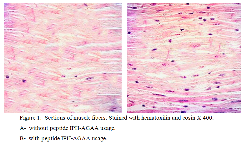
During the experiment, we found that in the control group in the quadriceps muscle’s section there were insignificant areas of fiber structuring, striation, which amounted to a total of 36.8±1.2% of muscle fiber restoration and indicates a high risk of contractures development and a decrease in muscle elasticity. While in rats, which were injected peptide IPH-AGAA, there is a structure of fibers, striations, and an area of the muscle fiber was 89.4±1.4%, and 2.4 times greater than without the use of the peptide IPH-AGAA (figure 1).

Thus, the peptide IPH-AGAA usage reduces the degree of inflammatory process in case of damage to muscle tissue, improves regeneration and restoration of muscle fibers, prevents the development of contractures and reduce muscle elasticity in the experimental model. These data indicate that the peptide IPH-AGAA usage optimizes metabolism in muscle cells, as well as has an antioxidant effect, prevents damage to muscle cells by free radicals during physical activity, also provides intensive and long-term nutrition of muscle cells, and has a stimulating effect on muscles in hypoxia and increases muscle flexibility and elasticity.
Conclusion
Thus, the peptide IPH-AGAA has an anti-inflammatory effect on muscle tissue damage and thus contributes to the improvement of regeneration of damaged muscles, which is manifested by cytostatic and anti-inflammatory action against muscle cells according to experimental studies.
The use of the peptide IPH-AGAA reduces the degree of inflammatory process in muscle tissue damage, improves regeneration and restoration of muscle fibers, prevents the development of contractures and reduce muscle elasticity in the experimental model. These data indicate that the use of the peptide IPH-AGAA optimizes the metabolism in the cells of muscle tissue, has an antioxidant effect, prevents damage to the cells of the neck tissue by free radicals, provides intensive and prolonged nutrition of muscle cells, stimulates the action on the muscles in hypoxia and increases the elasticity and resistance of muscles.
Literature
- Mironov A. N., Bunatyan N. D. etc. the Guidelines for preclinical studies of the medicine// the Team of authors. — M.: Grif and K, 2012. — 944 p.
- Khavinson V. Kh., Kuznik B. I., Linkova N. S., Pronyaeva V. E. The Influence of peptide regulators and cytokines on life expectancy and age-related changes in the hemostatic system // Advance of physiological Sciences. 2013. Vol. 44. № 1. P.39-53.
- Khavinson V. Kh. Peptide regulation of aging. : Science, 2009. — 50 p.
- Ahmetov II, Druzhevskaya AM, Astratenkova IV, Popov DV, Vinogradova OL, Rogozkin VA. The ACTN3 R577X polymorphism in Russian endurance athletes. Br J Sports Med. 2010 Jul;44(9):649-52.
- Kathryn R Wagner, MD, PhD and Julie S Cohen, ScM, CGC. Myostatin-Related Muscle Hypertrophy// National Center for Biotechnology Information, U.S. National Library of Medicine- 2018.- № 4- 35-39.
- Meienberg J, Rohrbach M, Neuenschwander S, Spanaus K, Giunta C, Alonso S, Arnold E, Henggeler C, Regenass S, Patrignani A, Azzarello-Burri S, Steiner B, Nygren AO, Carrel T, Steinmann B, Mátyás G. Hemizygous deletion of COL3A1, COL5A2, and MSTN causes a complex phenotype with aortic dissection: a lesson for and from true haploinsufficiency. Eur J Hum Genet. 2010;18:1315–21.


