The article presents the results of pre-clinical studies of the possibility of optimization of reparative osteogenesis in postoperative bone cavities of the jaws in old rats with a peptide bio regulator. Using the experimental study histological and morphometric methods, it is shown that the use of shot peptide positive effect on the course of the process of reparative osteogenesis of the mandible in old rats. The features of the medicine optimization of reparative osteogenesis jaw during aging. It is found that by using peptide in old animals to 30 days before surgery, the process of reparative osteogenesis standard defect of mandible occurs at early stages (before 30 days), similar to the control. On 60-120 days from the beginning of the experiment using the peptide bio regulators noted the formation of a complex regenerate tissue containing a smaller proportion of connective tissue and cartilage, as well as more advanced remodeling of newly formed bone tissue compared with the control group, and older animals who received the medicine after performing surgery.
Issues reparative age peculiarities of bone tissue regeneration are devoted to individual publications [1-3]. They indicate that with aging the quantitative and qualitative characteristics of proliferative process significantly decrease, which affects the severity of the recovery process in the healing of bone skeleton defects, namely, the restoration of bone tissue in the area of the former defect of old animals is characterized by a flaccid course and weak activation of stromal tissue along the periphery of the defect [4 — 6]. A number of scientific publications show age-related features of the course of reparative regeneration of the lower jaw bone tissue, as well as differences in the healing time of postoperative bone defects and the quality of regeneration in Mature and old rats [7 — 9]. In these works, it is noted that the process of reparative osteogenesis in the standard defect of the lower jaw, which arose after the removal of the left incisor, proceeds longer, with the formation of a complex tissue regenerate, a significant part of which is represented by Mature connective and cartilaginous tissues, remaining in the form of parts to the end of the experiment, which was completed on the 120th day after the creation of the jaw defect [10]. There are also few scientific works that consider the possibility of optimizing the reparative regeneration of jaw bone tissue during aging and the use of peptide bioregulators to solve this medical problem [11 — 13].
The aim of this experimental study is to examine the possibility of using the peptide bio-regulator to optimize the reparative regeneration of the bone tissue of the lower jaw in old rats.
MATERIAL AND METHODS
Experimental study on the possibility of introduction of peptide bio-regulator for optimization of reparative osteogenesis in older animals conducted on 45 white male rats of the Vistar line at the age of 22 — 24 months and weighing 400 — 650 g according to the method. Once rats were divided into 3 groups: control (postoperative bone cavity filled with blood clot) consists of 15 rats, First (experimental) group of 15 rats at the moment of the surgical intervention daily feed with added peptide (scheme 1) at the rate of 0.12 g per 10 g of body weight; Second (experimental) group of 15 rats for 30 days prior to the beginning of the experiment, daily feed with added peptide (scheme 2) at the rate of 0.12 g per 10 g of body weight.
The peptide bioregulator based on the Tripeptide, which has the ability to optimize the regeneration of bone and cartilage tissue, established in experimental and clinical studies [12].
Animals of all groups intraperitoneally hexenal anesthesia removed the lower left incisor. For that by a thin sharpened ironer, the gum was first peeled off and the periodontal tissues were crossed to a depth of 5 mm, and then by the hemostatic clip of the «mosquito» type, in which the shape of both cheeks of the working part of the tool was simulated in advance by thinning them and creating minor rounded depressions in them, the left lower cutter was removed. After removing the tooth, the edges of the gums above the hole were brought together by a seam, which allowed to reduce the gaping of the hole of the removed tooth and create the most optimal conditions for the formation of a blood clot, and subsequently to replace the bone tissue with newly formed tissues in the regeneration process.
Animals deduced from experience on 10, 20, 30, 60, 120-th day after surgery to remove the tooth. The preparation of the mandible was fixed in a 10% solution of neutral formalin, and then decalcified in solution using the GBI-1 device [7], carried out by ascending alcohols, poured into paraffin and prepared sections 5 microns thick, which were stained with hematoxylin and eosin by Van Gieson’s method. To be able to compare the results of the study with the data on the timing of reparative osteogenesis and bone regenerate composition in adult animals, the assessment of the course of the process of reparative osteogenesis in the hole of the removed tooth of rats of all groups was carried out using light microscopy and morphometry, according to the recommendations. At the same time, the percentage of bone, cartilage and connective tissue in the posttraumatic regenerate was estimated per unit area. Viewing and photographing of sections at various magnifications were performed in OPTON light-optical microscope at different magnifications of the lens and eyepiece. Also in this study, electron microscopy was used, for which the material was prepared according to the recommendations. All surgical interventions were carried out in compliance with the rules and requirements for equipment, instruments, aseptic and antiseptics, in accordance with the current «Rules of work using experimental animals». Statistical processing of the data was carried out using the Statistika 8.0 package (StatSoft, USA).
RESULTS AND DISCUSSION
A comparative study of histological preparations of the control and 1st experimental group of the study using light microscopy and morphometry showed some improvement in bone regeneration processes in group 1 compared with the control group. However, there were no significant differences in the course of reparative osteogenesis in the area of the removed tooth well at all times of the experiment (p>=0.05). Thus, the efficiency of application of peptide bio-regulator optimization of reparative osteogenesis when used according to scheme 1, whereby the start of injection is necessary on the first day of the experiment (day of surgery). At the same time, the appointment of peptide bio-regulator according to scheme 2 in the experimental group 2, when the peptide used for 30 days before the start of surgery on the lower jaw of an experimental animal obtained statistically significant results. In the 2nd experimental group, positive changes in bone tissue regeneration were noted in comparison with the control and the 1st experimental group, which were especially appeared on the 60-120 day from the beginning of the experiment and were characterized by both the tissue composition of the formed complex tissue regenerate, which contained a smaller proportion of connective and cartilaginous tissue, and more advanced processes of remodeling of the newly formed bone regenerate compared with the control and animals of the 2nd group. To be presented in a comparative perspective the dynamics of reparative osteogenesis in all experimental groups at the pre-clinical study of the efficacy of peptide on the reparative osteogenesis of the jaws.
The study of the healing bone defect of the jaw, formed after removal of the left lower incisor from older animals (control group) for 10-day of experiment has showed that the alveolus of the lower jaw of the filling jet changed young loose connective tissue, sometimes found isolated neutrophilic granulocytes, preserving the morphological features of its structure. Only in the bottom and surrounding walls of the alveoli showed signs of formation reticulations bone tissue.
A similar morphological pattern was obtained in the study of histological preparations obtained from animals of the 1st and 2nd experimental groups. In group 2, the analysis of the healing of the jaw bone defect on the 10th day of the experiment showed that the cavities, as well as in the control group, were filled with reactive altered loose connective tissue, in which single polymorphonuclear leukocytes and round cell elements were observed. In places along the edge and bottom of the defect (hole) it was possible to note the formation in the inward direction of the fiber reticulations bone tissue.
On the 20th day of the experiment in animals of the control group, as well as in animals of the 1st and Marked by the dynamics of the process of reparative osteogenesis in the hole of the tooth of the lower jaw in these groups remained at the 60th and 120th day of the experiment. After 60 days from the onset of the experiment, the animals of all 3 groups, the former the defect of the mandible, formed after removal of the left cutter, filled reticulations bone tissue with signs of its restructuring. In all histological preparations it was possible to clearly define several stages of maturation of newly formed bone tissue, namely remodeling of newly formed bone structures. At the same time a powerful beam reticulation tissue forming the osteons and lamellar bone tissue in a greater degree was detected in histological specimens obtained from the experimental animals of the 2nd group, which was confirmed by morphometric study. At the same time of the experiment, the animals of the control and the 1st experimental groups were detected starting the process of restructuring of the bone regenerate, with fabric, filling the former bone defect formed after removal of the lower left incisor, were still signs of a complex tissue regenerate.2nd experimental groups on the edge of the defect, active formation of reticulofibrous bone fiber exclusively along the edge of the defect was noted, although most of the former defect was still occupied by loose fibrous connective tissue. At this period of the study in specimens obtained from animals of all investigated groups identified fiber of the new formed reticulations bone tissue, which was defined most clearly at the periphery of the former defect (alveoli) and to a lesser extent in the Central part. Thus, on the 20th day of the experiment in all studied series of histological preparations the most active neoplasm of reticulofibrous bone tissue beams along the edge of the defect was revealed, especially in animals of the 1st and 2nd groups. The fiber of reticulofibrous bone tissue, regardless of the experiment series, were covered with numerous osteoblasts in the active state, as evidenced by their ultrastructural characteristics in the form of numerous cavities of the granular endoplasmic network filled with the content of moderate electron density.
The marked differences in the course of reparative regeneration of the jaw bone tissue have been clearly determined starting from the 30th day of the surgery, which was confirmed not only by the comparative morphological assessment of histological preparations, but also by the morphometric study data. On the 30th day of the experiment in all animals the well of the tooth was filled with a complex tissue regenerate. If in the control and the 1st group in the composition of the complex tissue regenerate connective tissue of different degrees of maturity prevailed, as well as parts of cartilage tissue, namely hyaline cartilage, then in animals of the 2nd group in the composition of the complex tissue regenerate, the proportion of reticulation tissue was significantly greater (^<0.05) than in animals of the control and 1st study groups. In addition, in animals of the 2nd group on the 30th day of the experiment, the beginning processes of bone regenerate restructuring and the single appearance of lamellar structures with good vascularization of a complex tissue regenerate that filled the cavity of the former jaw defect were noted.
This noted dynamics of the process of reparative osteogenesis in the hole of the tooth of the lower jaw in these groups remained at the 60th and 120th day of the experiment. After 60 days from the onset of the experiment, the animals of all 3 groups, the former the defect of the mandible, formed after removal of the left cutter, filled reticulations bone tissue with signs of its restructuring. In all histological preparations it was possible to clearly define several stages of maturation of newly formed bone tissue, namely remodeling of newly formed bone structures. At the same time a powerful beam reticulation tissue forming the osteons and lamellar bone tissue in a greater degree was detected in histological specimens obtained from the experimental animals of the 2nd group, which was confirmed by morphometric study. At the same time of the experiment, the animals of the control and the 1st experimental groups were detected starting the process of restructuring of the bone regenerate, with fabric, filling the former bone defect formed after removal of the lower left incisor, were still signs of a complex tissue regenerate.
After 120 days from the beginning of the experiment in animals of the 2nd group, the healing process of the bone defect can be considered almost complete. Tissue in the bone regenerate, filling the alveoli, the process of remodeling was virtually completed, the generated havers system, which is characteristic for lamellar bone tissue. For this period of study in animals of control and group 1, the former bone defect of the lower jaw, although filled with bone tissue, but the processes of its remodeling were significantly less advanced than in group 2. In the study of newly formed bone tissue in the control and 1st groups of animals maintained different stages of maturation of bone structures. Along with the emerging osteons and lamellar bone tissue, they retained fiber of reticulofibrous bone tissue, which indicates less advanced processes of bone regenerate remodeling. In some preparations in these groups of animals on the 120th day of the experiment it was possible to increase in the composition of the regenerate parts of hyaline cartilage, and in some places — Mature fibrous connective tissue, which testified to the preservation of heterogeneity of the developed regenerate in animals of the control and 1st experimental groups.
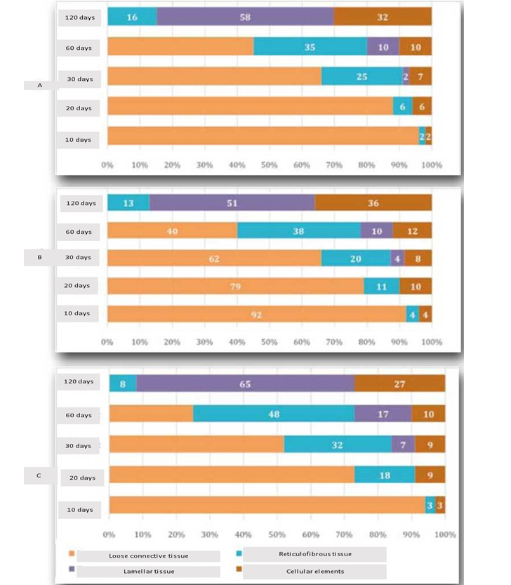
The dynamics of proliferation and bone cell ratio in regenerative osteogenesis of the mandible in rats in all study groups at different periods of the experiment allows to note that when using the shot peptide in old animals, the process of reparative osteogenesis in the standard defect of the mandible proceeds the same in the early stages (up to 30 days) and is similar to reparative osteogenesis occurring under a blood clot in the control group of animals. On the 60-120th day from the beginning of the experiment in the 2nd experimental group of animals with the use of peptide bio-regulator on the scheme 2 (30 days before surgery) noted the formation of a complex tissue regenerate containing a smaller proportion of connective and cartilaginous tissue, as well as more advanced processes of remodeling of newly formed bone tissue compared with the control and the 1st group of animals.
The figure shows the morphometric indicators of reparative osteogenesis in animals of 3 studied groups, which, as well as the data of morphological study, indicate that the use of the peptide, according to the scheme 2 allows to optimize the processes of reparative regeneration of the bone tissue of the lower jaw during aging in the experiment.
Thus, it can be argued that the histological and morphometric studies of the tissue composition of the regenerate in the hole of the removed left incisor of the mandible of old rats at different times of the experiment (from 10 to 120 days), as well as statistical analysis of the digital material obtained from the morphometry of bone Regenerated revealed the possibility of using the peptide to optimize reparative osteogenesis in aging. However, the optimization of bone tissue regeneration in the application of peptide was achieved only with a certain scheme of use of this medicine, which provides for long-term (1 month) its use in an experimental animal before surgery on the lower jaw.
CONCLUSION
The results of preclinical studies efficiency of application of shot peptide for optimization of reparative regeneration of bone tissue of the jaws during aging showed the possibility of using this medicine for these purposes. The study determined the method of the most effective use of this medicine to optimize reparative osteogenesis in aging — daily use of that for 30 days before surgery on the bone of the lower jaw of the experimental animal. It seems appropriate to test the use of peptide in clinical practice to optimize bone regeneration of the jaws in geriatric dentistry.
Figure below: Dynamics and proportion of the elements of the regenerate in the regeneration osteohistogenesis of the mandible of experimental animals of the control (A), First (B) and the Second (C) experimental groups in different periods of the experiment, unit (%).